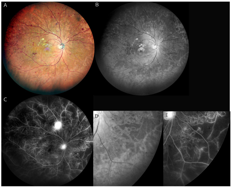
Figure 3.
(A): Multicolor widefield SLO image of the right fundus of a 65-year-old man with PDR. (B): Blue SLO image shows a hyporeflective area in the mid-periphery to periphery of the fundus, indicating non-perfused areas. (C): Widefield FA image shows widespread NPAs extensively with dye leakage from new vessels. (D): Magnified image of image (B) shows hyporeflective areas in the lower temporal quadrant. (E): Magnified FA image of image (C) shows NPAs in the same quadrant of image (D). All figures above are Mirante (NIDEK, Japan) images.

