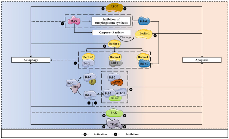
Figure 4.
The apoptosis/autophagy regulation loop. There are several apoptosis regulators that are also involved in the control of autophagy and both processes are often activated together to respond to stress stimuli. These interactions can result in both inhibition and induction of autophagy. To inhibit the autophagic process, Beclin-1 can bind to Bcl-2, Bcl-xL and Mcl-1. In contrast, under nutrient starvation conditions, Bcl-2 is prevented in binding to Beclin-1, after being phosphorylated by JNK1, allowing the initiation of autophagosomes formation. Furthermore, BAX can lead to Beclin-1 cleavage, which is dependent on caspase-3 activity. This pro-apoptotic protein can also inhibit the autophagosome synthesis, a process that can be reverted by Bcl-xL. In addition, activators of apoptosis, including TRAIL, can regulate autophagy, having a positive influence on this mechanism. Regarding apoptosis induction, calpain-cleaved ATG5 and Bcl-2 interaction facilitates this programmed cell death process. Moreover, ATG7 expression leads to autophagy induction, caspase activation and increased cell death. The conjugation ATG12-ATG3 can also regulate apoptosis, as its disruption may result in increased Bcl-2 expression and decreased apoptotic cell death.

