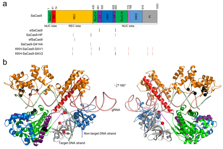
Figure 2.
The locations of high-fidelity SaCas9 mutations. (a) domain organization of SaCas9. The architecture by Nishimasu et al. [79] is used. The positions of the mutated residues for all high-fidelity variants are indicated by black vertical lines. The PAM-relaxed KKH mutations are shown in red vertical lines. (b) the high-fidelity point mutations, shown as black spheres, are mapped onto the structure of SaCas9 (PDB ID: 5AXW) [79]. The KKH mutations are shown as red spheres. The color scheme for the structure is identical to that for the sequence.

