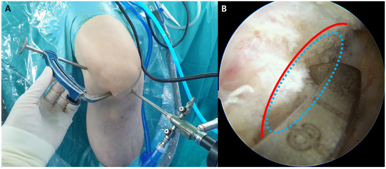
Figure 1.
Creation of an anatomical femoral tunnel using the outside-in technique. (A) After palpation of the lateral femoral epicondyle, a small incision is made just proximal and posterior to the lateral femoral epicondyle. The guide’s angulation is adjusted to allow the tip of drill sleeve placement in the incision at the femoral footprint of the anterolateral ligament. (B) The tip of the femoral aiming guide is placed at the femoral footprint of the anterior cruciate ligament through the anterolateral portal under direct arthroscopic visualization through the anteromedial portal (red line, lateral intercondylar ridge (Resident’s ridge)); blue ellipse, femoral footprint of the anterior cruciate ligament).

