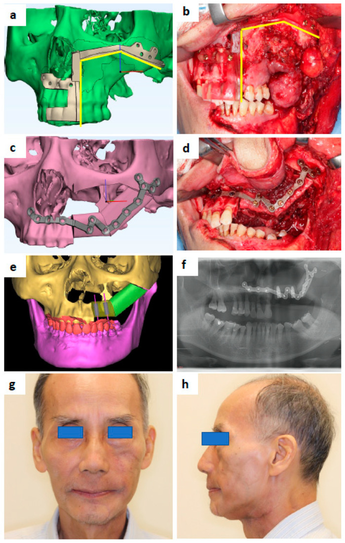
Figure 1.
A 69-year-old male that presented with carcinoma ex pleomorphic adenoma at the left maxilla. (a) Maxilla resection guide design. (b) 3D-printed maxilla resection guide fitted intraoperatively. (c) Patient-specific Titanium plate design. (d) 3D-printed Ti plate fitted intraoperatively. (e) Design showing the location of simultaneous dental implants to be inserted during fibula free flap harvest. (f) Post-operative orthopantomography. (g) Postoperative 7 months and post-radiation 4 months—frontal view. (h) Profile view.

