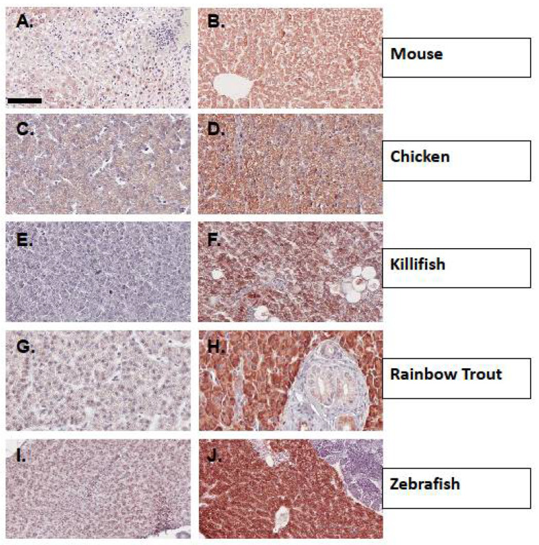
Figure 3.
Immunohistochemical detection of CYP1A protein expression using mAb CRC4 in liver tissues from representative vertebrates. (A). Control mouse liver; (B). Liver from a PCB-126 treated mouse; (C). Control chicken embryo liver at 18 days post fertilization (DPF); (D). Liver from a crude oil vapor-exposed chicken embryo at 18 DPF; (E). Liver from CYP1A recalcitrant Gulf killifish; (F). Liver from Gulf killifish collected from a crude oil exposed site; (G). Control Rainbow trout liver; (H). Liver from a β-NF-exposed rainbow trout; (I). Control zebrafish liver; (J). Liver from PCB-126 treated zebrafish. (A,B) are 20× magnification, scale bar = 100 µm (C–J) are 40× magnification, scale bar = 50 µm. Details of tissue sources, experimental conditions, and procedures are found in Methods and Table 2.

