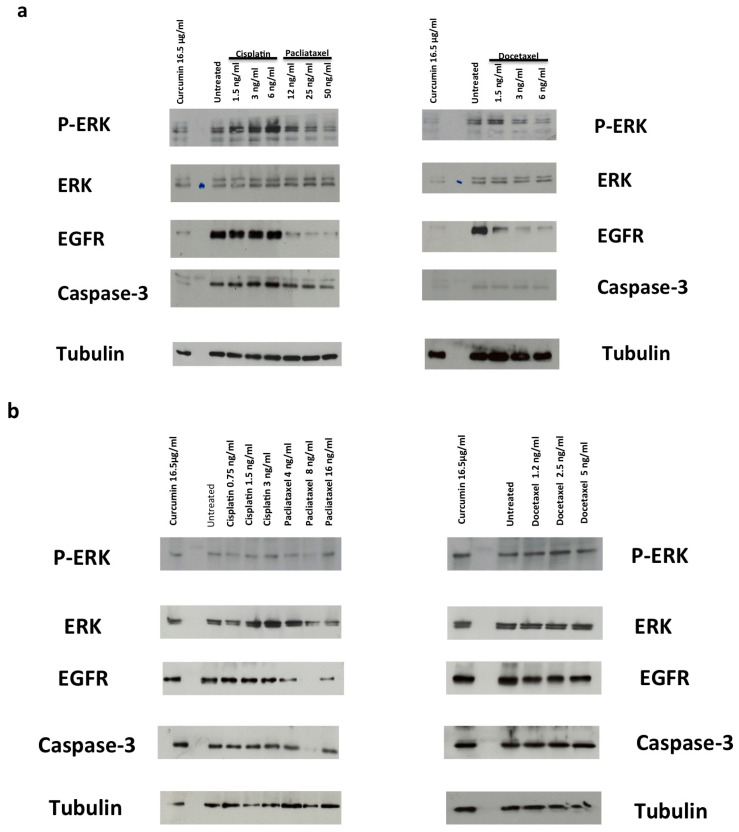
Figure 5.
(a,b) Role of the p-ERK/ERK/EGFR in survival and apoptosis. DU-145 (a) and PC-3 (b) cells were used. In (a,b), the cells were stimulated for 48 h in the absence or presence of the indicated compounds. Curcumin was used at 16.5 µg/mL (44.79 µM). Proteins were separated by SDS-PAGE and then analyzed by Western blot, using the antibodies against the indicated proteins.

