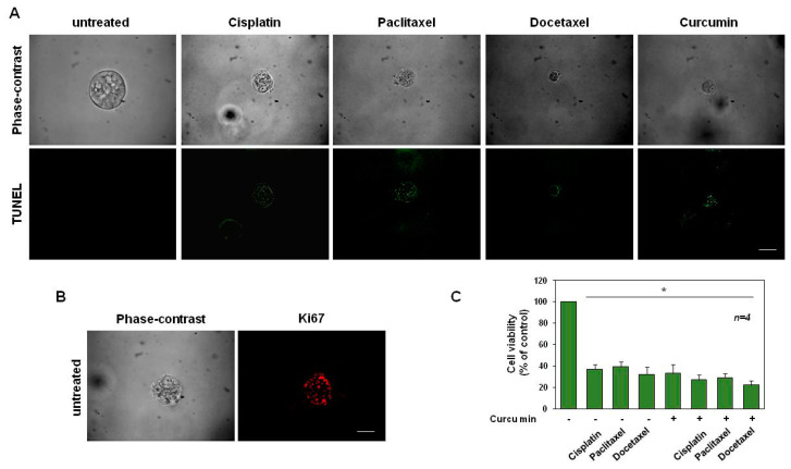
Figure 8.
Cisplatin, paclitaxel and docetaxel (used at 25 ng/mL) and curcumin (used at 6.9 µg/mL–18.73 µM) induce apoptosis in DU145 cells. (A): Phase-contrast and in situ cell death KIT-stained images (green) of organoids left untreated or treated in the absence or presence of the indicated drugs for 15 days are shown. Scale bar, 100 µ. (B): Phase-contrast and Ki-67-stained images (red) of organoids untreated for 15 days are shown. Scale bar, 100 µ. In (A,B), images are representative of three different experiments, each in duplicate. (C): Cell viability was evaluated by MTT assay in DU145 organoids after 12 days of culture. The graph in (C), represents the organoids’ cell viability expressed as % of control, assumed as 100% of viability. Four independent experiments were carried out. Means and standard error of the means (SEMs) are shown, and “n” represents the number of the experiments. *: p < 0.05 for the indicated experimental points vs. the untreated control.

