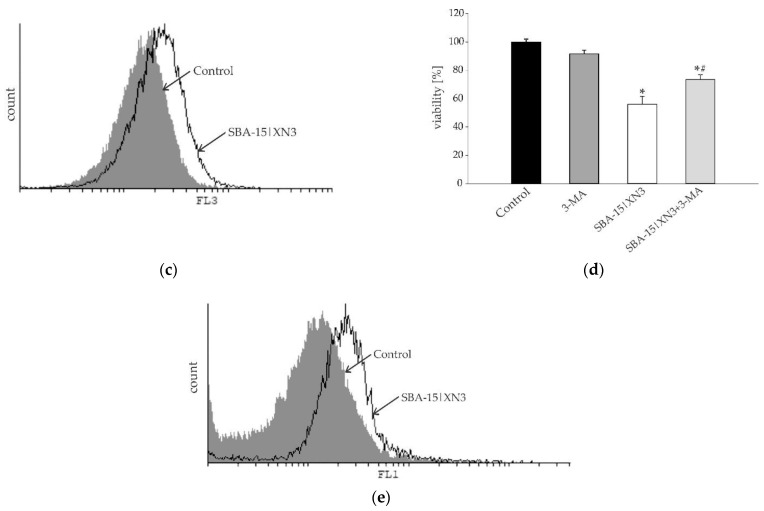
Figure 5.
Inhibition of cellular proliferation and promotion of autophagy without induction of caspase-dependent apoptosis in B16F10 melanoma cell culture determined using Ann/PI (a), ApoStat (b), AO (c) and CFSE (e) staining, all performed after 48 h of treatment with SBA-15|XN3 and subsequently analyzed by flow cytometry. Dot plots and histograms are representative ones selected from three repeated experiments. The induction of autophagic cell death exposed to MC50 dose SBA-15|XN3 and 3-MA (in final concentration of 1 mM) for 48 h (d). Cell viability was determined by CV assay and expressed as a percentage of control values (untreated cells). The data are presented as mean ± SD obtained from three independent experiments. * p < 0.05 refers to untreated cultures; # p < 0.05 refers to SBA-15|XN3 treated cultures.


