Figure 3.
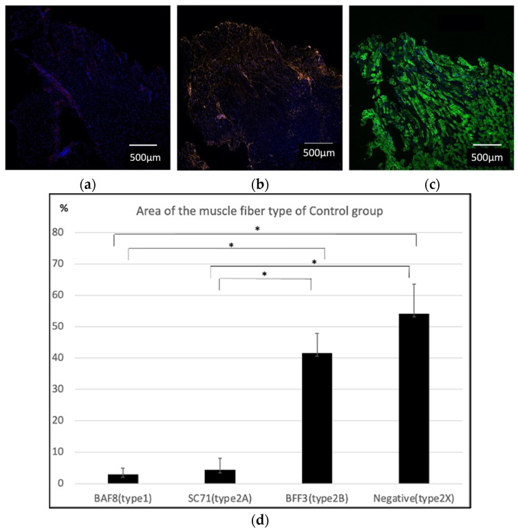
Immunohistochemistry of the masseter muscle in the control group. (a) Anti-BAF8 (red) was used for the staining of type 1 muscle fibers, and almost all specimens were negative. (b) Anti-SC71 (orange) staining was performed to detect type 2A fibers, and a slightly positive area was observed. (c) Anti-BFF3 (green) staining suggested that most of the masseter muscle fibers were composed of type 2B fibers. Nuclear counterstaining was performed using DAPI (blue). (d) Anti-BAF8-, anti-AC71-, and anti-BFF3-positive areas were analyzed with four specimens. The average percentage area of type 1, type 2A, type 2B and the estimated type 2X fibers were 2.92 ± 2%, 4.40 ± 3.62%, 41.52 ± 6.30% and 51.16 ± 6.77%, respectively. This indicated that the original masseter muscle was composed mainly of type 2 fibers, especially type 2B fast-twitch glycolytic muscle fibers (n = 4 each, * p < 0.01, one-way ANOVA).

