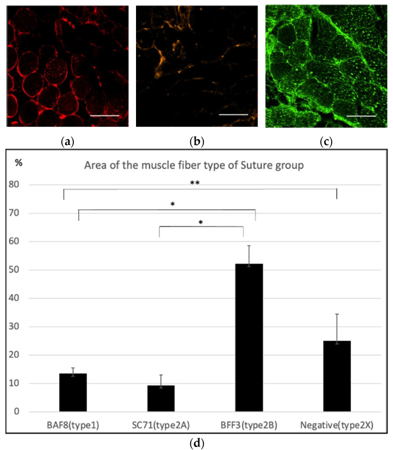
Figure 4.
Immunohistochemistry of the masseter muscle in the suture group. (a) Anti-BAF8 (red) staining was slightly positive in some areas. (b) Anti-SC71 (orange) staining was performed to detect type 2A fibers, and a slightly positive area was observed. (c) Anti-BFF3 (green) staining suggested that most of the masseter muscle fibers in the suture group were composed of type 2B fibers. Nuclear counterstaining was performed using DAPI (blue). Scale bar = 100 µm. (d) Anti-BAF8-, anti-SC71-, and anti-BFF3-positive areas and the negative area for all antibodies were analyzed with four specimens. The average percentage area of type 1, type 2A, type 2B and the estimated type 2X fibers were 13.50 ± 2.83%, 9.38 ± 2.01%, 52.17 ± 8.30% and 25.00 ± 7.72%, respectively. (n = 4 for each group, * p < 0.01, ** p < 0.05, one-way ANOVA).

