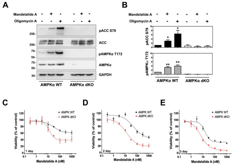
Figure 3.
Mandelalide-induced phosphorylation of ACC requires AMPK and confers a survival advantage to wild-type mouse embryonic fibroblasts (MEFs). (A) Immunoblot analysis of wild-type and AMPKα-null double knockout (dKO) MEFs treated with mandelalide A (30 nM) or oligomycin A (1 µM) for 90 min. Whole cell lysates were probed with antibodies for phospho-AMPK, phospho-ACC, total AMPK, total ACC and GAPDH as indicated. Blots are representative of a single experiment that was repeated three times (B) Histograms show quantification of immunoblot data shown in A from three independent experiments. Values represent band intensity of phospho-AMPK/total AMPK and phospho-ACC/total ACC, normalized to GAPDH. Statistical significance of change relative to vehicle (0.1% DMSO) is indicated as * p < 0.05, ** p < 0.01. (C–E) Concentration-dependent change in the viability of AMPKα-null MEFs and wild-type MEFs after 24 h (C), 48 h (D) and 72 h (E). MEFs were continuously exposed to mandelalide A or vehicle (0.1% DMSO) and cell viability determined at the endpoint of the assay using a CellTiter-Glo® assay with the viability of vehicle-treated cells defined as 100%. Graphs represent mean viability ± S.E. (n = 3 wells per treatment) and curves represent the fit of data points by nonlinear regression analysis to a logistic equation. Data represent one comparison from at least three independent experiments.

