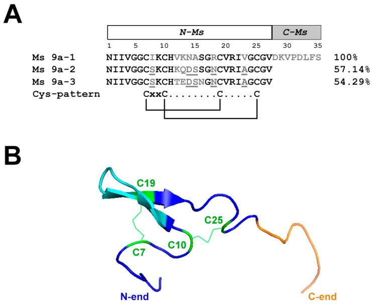
Figure 1.
Alignment of Ms 9a homologues and spatial structure of Ms 9a-1. (A). Comparison of the amino acid sequences of Ms 9a-1, Ms 9a-2, and Ms 9a-3. Similar for all three peptides residues marked in black, variable regions marked in grey, mismatches in these regions underlined. Amino acids of Ms 9a-1 from 1 to 27 correspond to the engineered peptide N-Ms, while residues from 28 to 35 are C-Ms. (B). Spatial structure model of Ms 9a-1. Cys residues are green, the variable region between C10 and C19 is cyan, and non-homologous C-tail (С-Ms) is orange.

