
Figure 2.
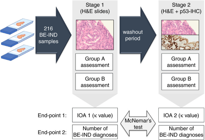
Design of the study. In stage I, each slide was assessed by one pathologist from group A and one pathologist from group B. After a wash‐out period of at least 8 weeks, the slides were assessed with matched p53‐stained slides available by the same pathologists as in stage I. In stage II, the pathologists were blinded to their original diagnosis. BE‐IND, Barrett's oesophagus indefinite for dysplasia; H&E, haematoxylin and eosin (staining); IOA, interobserver agreement. P53‐IHC, P53 immunohistochemistry.