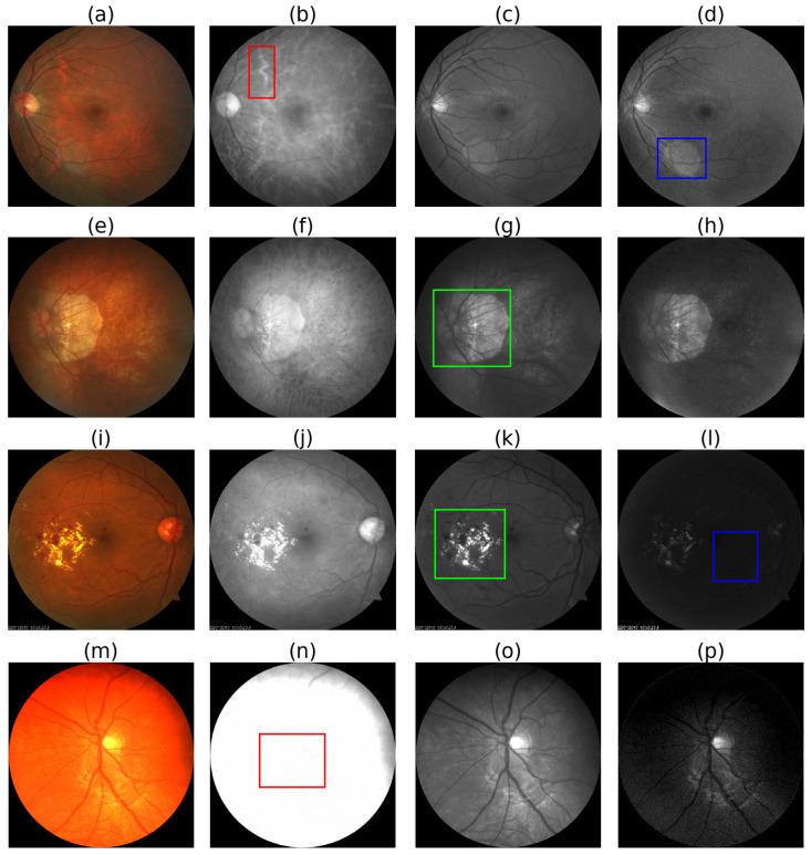
Figure 3.
Pros and cons of different color channels. 1st column i.e., (a,e,i,m): RGB fundus photographs, 2nd column i.e., (b,f,j,n): red channel images, 3rd column i.e., (c,g,k,o): green channel images, and 4th column i.e., (d,h,l,p): blue channel images. Choroidal blood vessels are clearly visible in the red channel, as shown inside the red box in (b). Lens flares are more visible in the blue channel, as shown inside the blue box in (d). Atrophy and diabetic retinopathy affected areas are more clearly visible in the green channel as shown inside the green boxes in (g,k). As shown inside the blue box in (l), the blue channel is prone to underexposure. The red channel is prone to overexposure, as shown inside the red box in (m). Source of fundus photographs: (a) PALM/PALM-Training400/H0025.jpg, (e) PALM/PALM-Training400/P0010.jpg, (i) UoA_DR/94/94.jpg, and (m) CHASE_DB1/images/Image_11L.jpg.

