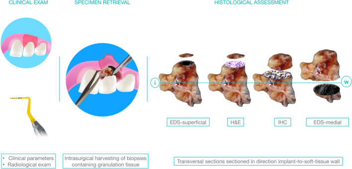
FIGURE 1.

Study design. Diagnostic specimen represented granulation tissue harvested within routine surgical treatment of peri‐implantitis/periodontitis using Ti‐free instruments. In order to ensure true‐positive outcomes, the elemental analysis was performed using scanning electron microscopy (SEM) and dispersive X‐ray spectrometry (EDS) in the first and last (medial) sections following consecutive sectioning for haematoxylin and eosin staining (H&E), and immunostaining. Histopathological assessment was performed in H&E sections, while the nuclear factor kappa‐B (Nf‐kB), interleukin‐6 (IL‐6), Cluster of Differentiation 68 (CD68) and vascular endothelial growth factor (VEGF) were used to compare inflammatory patterns between the groups
