FIGURE 2.

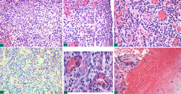
Histopathological profile in peri‐implantitis. Inflammatory infiltrate with focal presence of neutrophils was observed in all samples suggesting the infection‐induced chronic inflammation as a common pathological diagnosis of peri‐implantitis. Two patterns of inflammation were observed in PI, including chronic lymphocyte infiltrate with abundant plasma cells (a) and subacute infiltrate (b) characterized with chronic infiltrate interweaved with zones of neutrophil (N) granulocytes and sporadic eosinophils (E) (violet arrows indicate pink zones of granulocyte infiltrates within chronic infiltrate). Neutrophils were observed in all specimens in the lining contact zone towards dental implant (c, green arrows), the squeezed neutrophils were observed in the endothelium and vessel lumen (d) as well as in the extravasated blood content (e, f). Peri‐implantitis lesions exerted dense vascular networks with hyperaemic vessels that were frequently associated with micro‐bleeding characterized with focal erythrocyte infiltrates (f). All micrographs have been captured at x40 magnification, while the d and e represent zoomed crops of the same micrographs aiming at better visualization of specific histopathological details
