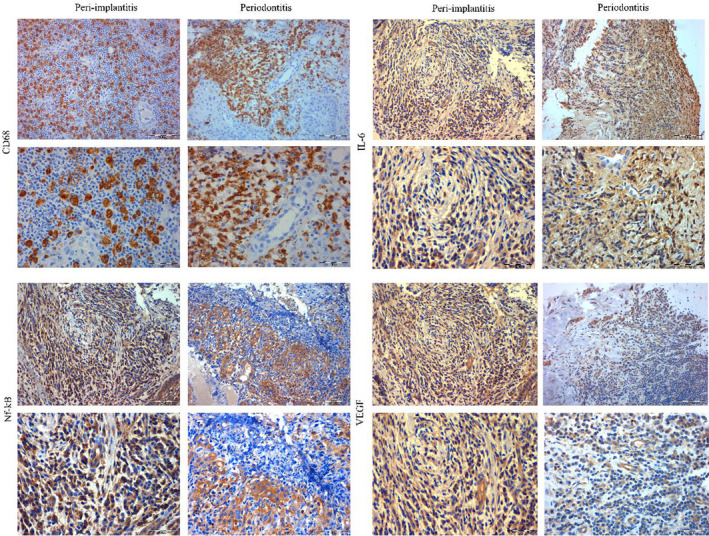
FIGURE 3.

Expression of immunohistochemical markers between peri‐implantitis and periodontitis. Peri‐implantitis and periodontitis granulation tissue exhibited intensive staining for CD68, Nf‐kB and IL‐6, while the CD68 was the single marker significantly more expressed in peri‐implantitis. VEGF was significantly more expressed in peri‐implantitis when compared to periodontitis where it was lightly expressed. Micrographs were captured at two magnifications x200 and x400 per each experimental and control sample
