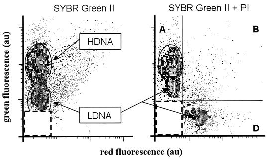
FIG. 7.
Comparison of single (SYBR Green II; 1:1,000 [vol/vol] final dilution) and double (NADS protocol) staining of bacteria in a seawater sample collected off Marseilles at the site facing Maire Island. The green versus the red fluorescence cytograms corresponding to SYBR Green II staining reveal the presence of bacteria with high (H) and low (L) DNA contents. The cytogram associated with the NADS protocol shows a significant amount of LDNA bacteria appearing as red-negative cells in quadrant A. At the same time, cells that were negative with SYBR Green II were apparent with PI staining in quadrant D. Refer to Table 1 for the counts.

