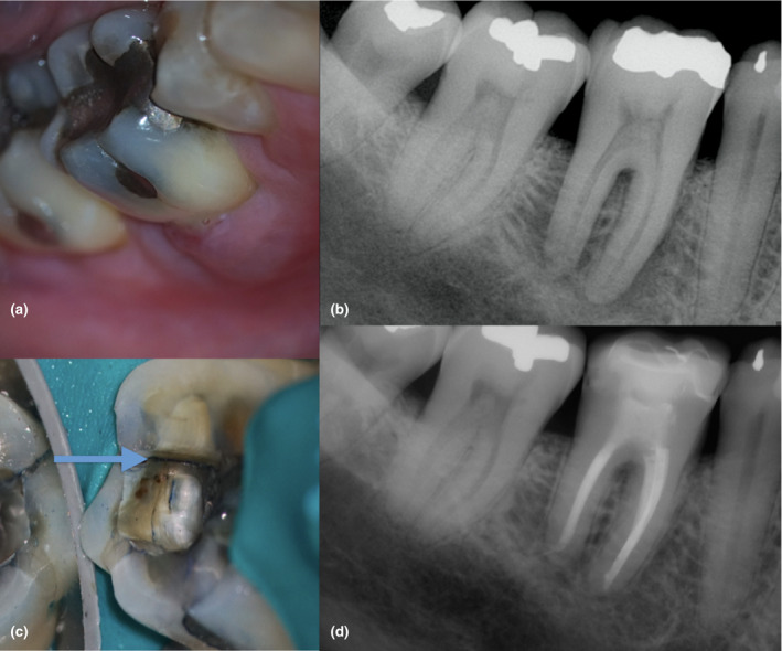
FIGURE 10.

Mandibular first molar with pulp necrosis, chronic apical abscess, and furcation involvement in an immunocompetent older adult (a). The periapical digital image shows a radiopaque restoration consistent with an amalgam occlusal filling in the mandibular first, second and third molar, an ill‐defined radiolucency with a lateral root lesion, and furcation involvement associated with #30 (b). Observe the presence of a crack in the distal wall (c). Three prognostic factors were identified in this case: presence of a distal crack, periapical pathosis and probing depth higher than 4 mm (Krell & Caplan, 2018). Prognosis was unfavourable. The treatment plan option accepted by the patient was debridement, placement of calcium hydroxide medication and a temporary crown in the affected molar. The case was completed 3 months later after initial signs of healing were confirmed. Despite the unfavourable prognosis, the 18‐month follow‐up radiograph shows significant healing (d). The case was classified as healing
