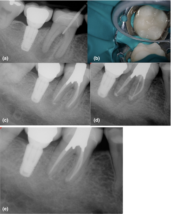
FIGURE 12.

Mandibular first molar with pulp necrosis and chronic apical abscess in an immunocompetent older adult (a). Observe the J‐shaped lesion surrounding the distal root, furcation involvement and a calcified pulp chamber. The prognosis was questionable. The presence of an indirect restoration did not avoid a conservative treatment (b). The case was completed after the sinus tract healed and probing depth was <4 mm. A two‐visit model was used to manage this chronic infection (c‐d). The 12‐month recall shows satisfactory healing of the furcation area and periapical tissues. Signs of healing avoided a future surgical intervention in this case with complex bone loss pattern and a possible apico‐marginal defect (e)
