
FIGURE 14.
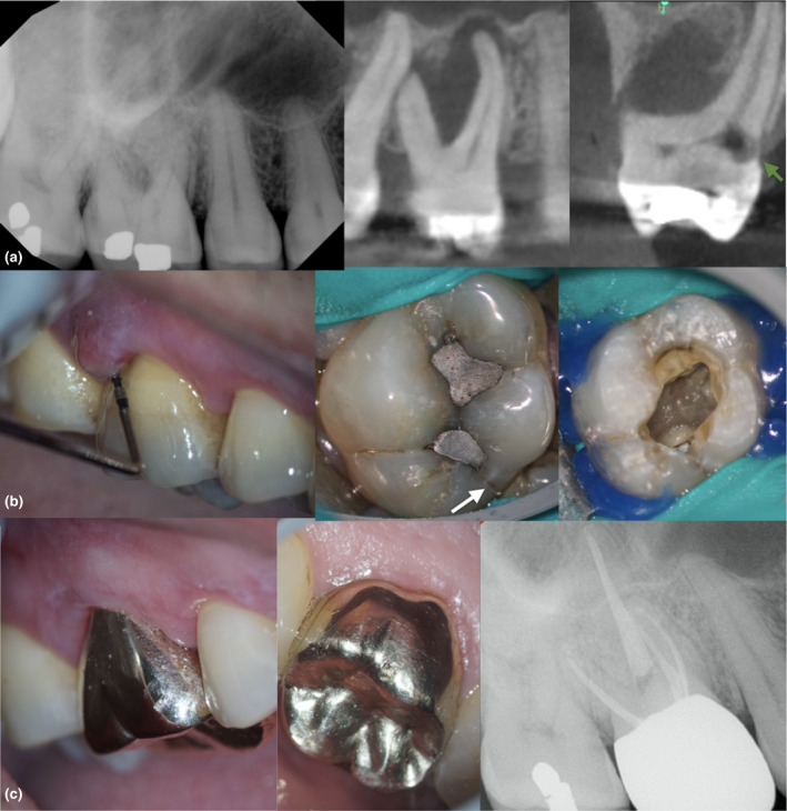
Fifty‐six‐year‐old patient with a chronic apical abscess and pulp necrosis in a maxillary first molar. No systemic conditions were recorded. The periapical image shows the presence of periapical radiolucencies and furcation involvement. Limited field‐of‐view CBCT sagittal and coronal sections revealed the presence of an extensive furcation defect, a lateral root lesion and external invasive cervical resorption (EICR) in the palatal side (a–b). The clinical inspection revealed the presence of a crack in the distal marginal ridge associated with occlusal amalgam restorations. The inspection of the pulp floor revealed that the crack did not extend below the CEJ level. The prognosis was unfavourable because of the presence of several factors including extensive bone loss, EICR and a crack in the distal marginal ridge. The treatment plan options included extraction, debridement and intracanal medication or no treatment. Patient elected the non‐surgical treatment option, including the use of intracanal dressing. The common understanding between the provider and the patient was that the treatment will be completed after showing initial signs of healing (decrease in probing depth) and closure of the sinus tract. Occlusion was reduced after treatment, and a full‐coverage restoration was recommended after 3 months of follow‐ups. (c) The 18‐month recall shows that the apical tissues are healing