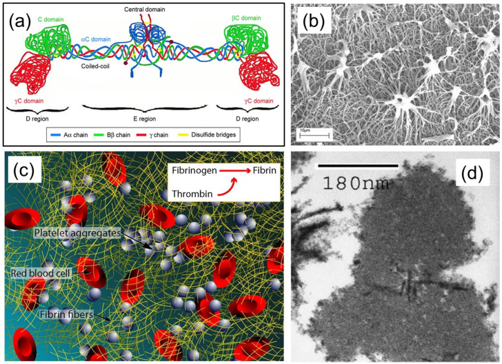
Figure 1.
(a) Fibrinogen structure. Aα chains are shown in blue, Bβ chains are shown in green, and γ chains are shown in red. Disulfide bridges stabilizing the coiled-coil regions are shown in yellow. Reprinted/adapted with permission from Ref. [5], 2017, DovePress. (b) SEM image of fibrin clots formed using high and low thrombin concentrations after 10 min of lysis after the addition of tissue-type plasminogen activator (tPA) to the surface of the clot. Reprinted/adapted with permission from Ref. [6], 2011, American Society of Hematology. (c) Image of how fibrin fibers from fibrinogen act as glue and a scaffold for the grouping of red blood cells, platelets, and other plasma proteins within a fibrin clot. Reprinted/adapted with permission from Ref. [7], 2017, Elsevier. (d) TEM image (90,000×) of the cross-section of a fibrin fiber. Reprinted/adapted with permission from Ref. [8], 2004, Elsevier.

