Figure 10.
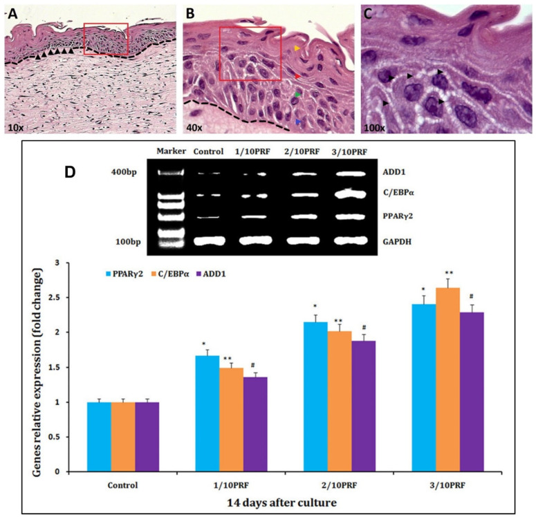
Histological features of skin organotypic cultures (ORGs) determined by H&E, after 21 days. (A) Whole structure of the ORGs (10×). A clear dermal–epidermal separation (dashed line) and a possible basal membrane (black head arrows) can be seen. (B) The enhanced area (40×) indicated by the red inset in (A). Four differentiation stages of the epidermis: the basal layer (blue head arrow), the spinous layer (green head arrow), the granular layer (red head arrow), and the horny layer (yellow head arrow), and the morphological associated changes in keratinocytes through the different layers of the epidermis. (C) Enhanced (100×) image indicated by the red inset in (B). The black arrows indicate hemidesmosome-like structures between keratinocytes. (A–C) Reprinted/adapted with permission from Ref. [97], 2016, Elsevier. (D) mRNA levels of PPARγ2, C/EBPα, and ADD1 mRNA, which are adipogenic marker genes, are higher in the platelet-rich fibrin (PRF) groups than in the control group after 14 days of culture. * p < 0.01, ** p < 0.01, # p < 0.01. Reprinted/adapted with permission from Ref. [98], 2017, Impact Journals, LLC.

