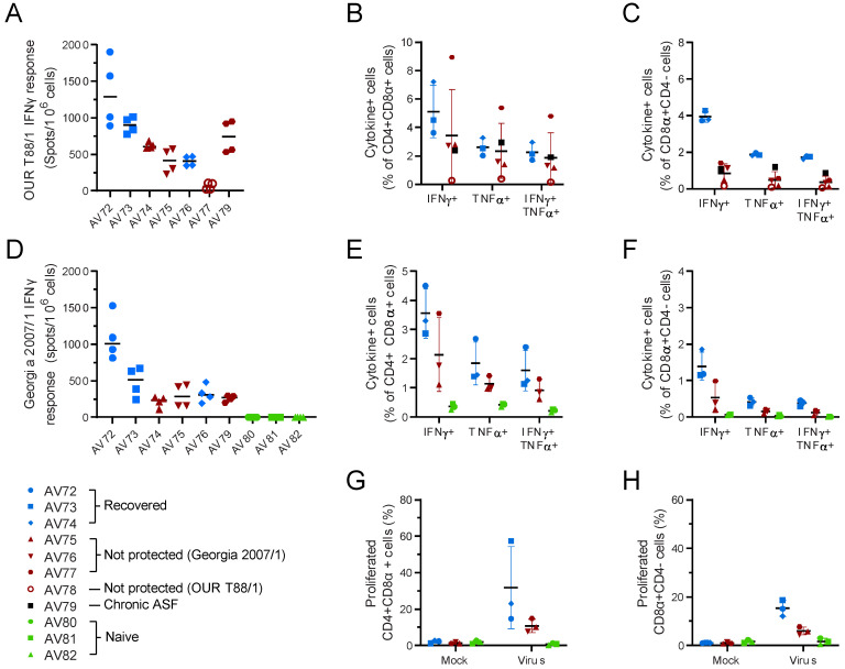
Figure 10.
Cellular responses to ASFV in outbred pigs. Animals were inoculated with low virulent ASFV isolate OUR T88/3 and then challenged twenty-one days later with highly virulent OUR T88/1, followed by Georgia 2007/1 twenty-one days after that. Blood samples were taken prior to challenge with OUR T88/1 on day 21 (A–C) or Georgia 2007/1 on day 42 (D–H). IFNγ secreting cells were enumerated by ELISpot (A,D) or flow cytometry (B,C,E,F) after stimulation with OUR T88/1 (A–C) or Georgia 2007/1 (D–F). The proportion of CD4+CD8α+ (B,E) or CD8α + CD4- cells expressing IFNγ, TNFα or both cytokines was determined using ICS. PBMCs purified before Georgia 2007/1 challenge were stimulated for 6 days with Georgia 2007/1 or a mock inoculum and the proportion of CD4+CD8α+ (G) and CD8α+CD4- (H) cells proliferating after 6 days stimulation with Georgia 2007/1 identified. Animals that recovered after challenge with Georgia 2007/1 are indicated in blue and those that did not in dark red. AV77 that did not recover after OUR T88/1 challenge is indicated as an open red circle and AV78 that suffered chronic ASF as a black square. Lines indicate the mean, and the error bars indicate the standard deviation from that mean.

