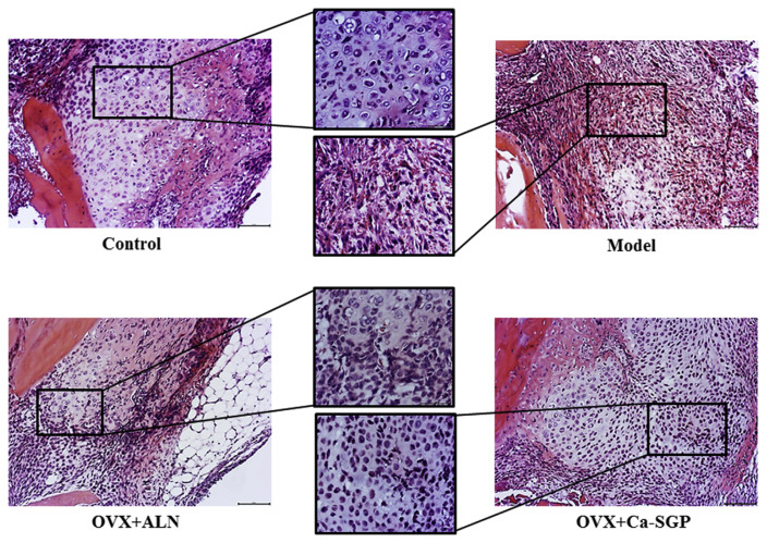
Fig. 3.
H&E staining of the callus on day 5 post-surgery. The calluses were fixed in 10% neutral formaldehyde for 24 h and then decalcified in 10% EDTA for 3 weeks. The decalcified tissues were embedded in paraffin after dehydration with ascending grades of ethanol. Then, the embedded samples were serially sectioned (thickness = 4 μm) and stained with H&E. Finally, the tissue slices were observed and photographed using BH-2 microscope equipped with matching camera software (n = 6, 10 × and 20 × magnification).

