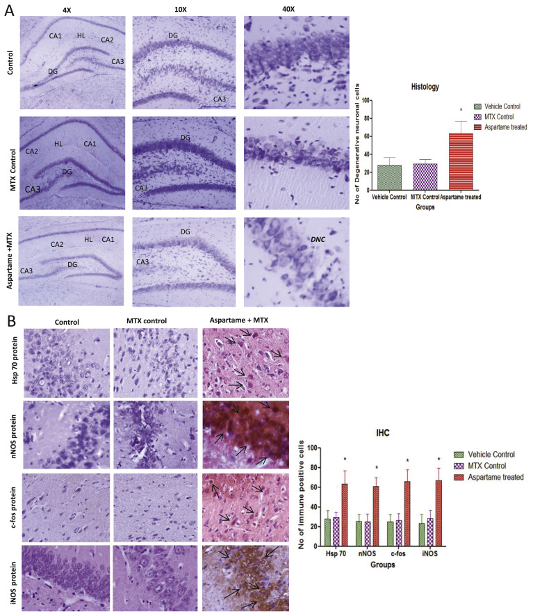
Fig. 5.
Effect of long term aspartame (40 mg/kg b.wt) on brain in Wistar albino rat, [A] the histomicrograph of brain stained by cresyl fast violet. [B] Imunohistochemistry of Hsp70, nNOS, c-fos and iNOS protein expression in brain of Wistar albino rats. [A] Histomicrograph Cresyl fast violet (CFV) staining of brain in Control, Methotrexate (MTX) control and Aspartame + Methotrexate treated animals. CA1 – Cornus Ammonis 1, CA2 – Cornus Ammonis 2, CA3 – Cornus Ammonis 3, DG – Dentate Gyrus, DNC – Degenerative neuronal cells (DNC), HL – Hippocampal Layer. [B] Immunohistomicrograph staining of brain in Control, Methotrexate (MTX) control and Aspartame + Methotrexate treated animals. Comparison and analysis were done by the one-way analysis of variance (ANOVA), control group was compared with MTX control group and aspartame MTX group, MTX control group was compared with Aspartame MTX group. Data are expressed as mean ± SD, n = 6. *P ≤ 0.05. Aspartame treated group when compared to control significance is marked as * and MTX treated groups significance is marked as #.

