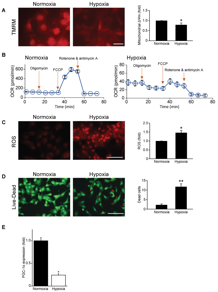
Figure 1.
Mitochondrial function, respiration, and cell viability are impaired in hypoxic cardiac myocytes. (A) Mitochondrial membrane potential (∆Ψ m) during normoxia and hypoxia as assessed by tetra-methylrhodamine methylester perchlorate (TMRM) staining (red fluorescence); bar: 20 μm. (B) Mitochondrial oxygen consumption rate (OCR) in neonatal rat cardiac myocytes subjected to normoxia (left panel) and hypoxia (right panel) from 6 to 8 wells per condition and from 3 to 4 independent myocyte isolations. (C) Mitochondrial reactive oxygen species (ROS) during normoxia and hypoxia as assessed by dihydroethidium staining (red fluorescence); bar: 100 μm. (D) Cell viability during normoxia and hypoxia. Green cells indicate live cells and red cells indicate dead cells; bar: 100 μm. (E) PGC-1α mRNA expression under normoxic and hypoxic conditions; mRNA levels were normalized to Gapdh. Data are expressed as mean ± SEM. * p < 0.05 or ** p < 0.01 versus normoxia, n = 3–4 independent myocyte isolations.

