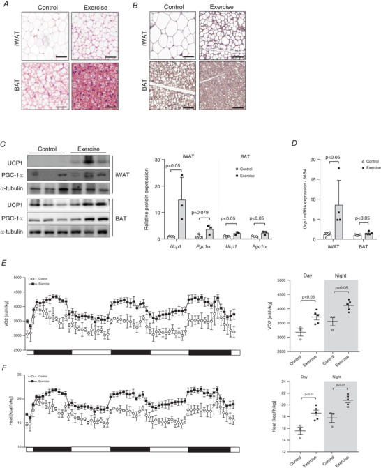
Figure 2. Voluntary wheel‐running exercise training induces browning of inguinal white adipose tissue (iWAT) and activation of brown adipose tissue (BAT).

A, haematoxylin and eosin (H&E) staining of iWAT and BAT tissues, n = 3 for all groups. B, immunohistochemical staining of iWAT and BAT for detection of uncoupled protein‐1 (UCP1) levels, n = 3 for all groups (scale bar = 50 μm). C, UCP1 and peroxisome proliferator‐activated receptor‐gamma coactivator‐1 alpha (PGC‐1α) protein expressions in iWAT and BAT, n = 3 for all groups (scale bar = 100 μm); quantification of protein expression, n = 3 for all groups. D, Ucp1 gene expression in iWAT and BAT, control (CON) = 5, exercise (EX) = 4. The expression levels were normalized to the expression of 36B4 gene. E, whole‐body oxygen consumption rate (VO2) (ml/h/kg) were monitored in mice, CON = 3, EX = 5. F, whole‐body heat generation (kcal/h/kg) was monitored in mice, CON = 3, EX = 5. Data are represented as means ± SD. Differences between two groups were analysed using a two‐tailed t test. [Colour figure can be viewed at wileyonlinelibrary.com]
