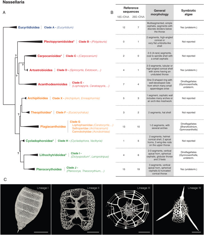
Fig. 5.

Schematic morpho‐molecular classification of Nassellaria based on ribosomal genes (18S and 28S rDNA) from Sandin et al. (2019).
A. Phylogeny showing molecular clades, taxonomic super‐families. Group with an asterisk denotes changes in taxonomic nomenclature from Sandin et al. (2019) following the latest classification scheme for Radiolaria (Suzuki et al., 2021). Non‐supported nodes are reported by a surrounded question mark. Colours correspond to molecular lineages (ML): blue = ML I, red = ML II, orange = ML III and green = ML IV.
B. Corresponding extent of the morpho‐molecular framework and main morphological features (including the presence and type of symbionts reported).
C. Scanning electron microscopy illustrations of typical silicified structures found in the four main nasellarian molecular lineages. All scale bars are 50 μm. Images courtesy of Miguel Sandin (Uppsala University).
