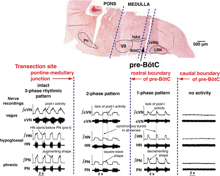
Figure 3. Hierarchical rostro‐caudal spatial organisation of the brainstem respiratory network.

Top, a sagittal section of the brainstem after transverse serial transections (note cut lines). Dashed lines indicate the transections at the pontine–medullary junction, the rostral boundary of the pre‐BötC, and the caudal boundary of the pre‐BötC. Bottom, the respiratory rhythm recorded from the central end of the vagus nerve (cVN; inspiratory and post‐inspiratory discharge), the hypoglossal nerve (HN; pre‐inspiratory and inspiratory) and the phrenic nerve (PN; inspiratory ramp) before (three‐phase) and after (two‐phase) ponto‐medullary transection. The resulting two‐phase pattern has no post‐inspiratory or pre‐inspiratory activities. The rhythm that exists with a transection at the rostral extent of the pre‐BötC is a single‐phase decrementing inspiratory pattern that is phase locked across all motor outputs and is abolished with sectioning at the caudal end of the pre‐BötC. Abbreviations: LRN, lateral reticular nucleus; NAd, nucleus ambiguous; Pn, pontine nucleus; rVGRG, rostral ventral respiratory group; VII, facial nucleus. Data from Smith et al. (2007).
