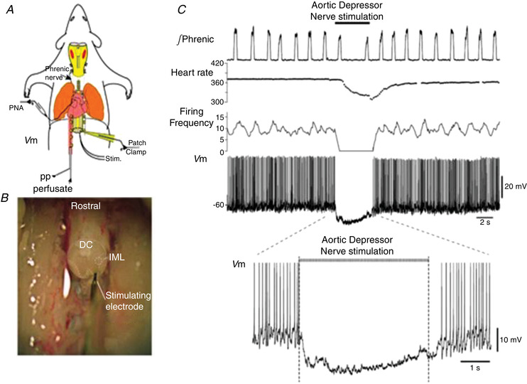
Figure 6. Whole cell recordings of functionally identified pre‐ganglionic sympathetic recordings in neonatal rats.

A, schematic WHBP of neonatal rat. Phrenic nerve and patch clamp recordings from sympathetic pre‐ganglionic neurones (SPN) were made. B, thoracic spinal cord is exposed and an oblique cut through the cord was made to visualise inter‐mediolateral (IML) cell column. SPNs were antidromically activated by stimulating the ventral root. DC, dorsal columns. C, electrical stimulation of the aortic depressor nerve hyperpolarised SPNs and caused a marked bradycardia. This baroreceptor inhibition was of short latency (within 100 ms). Data from Stalbovskiy et al. (2014).
