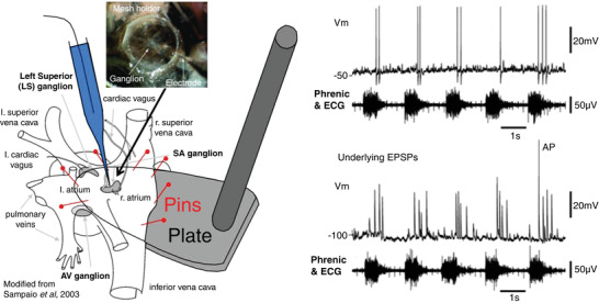
Figure 9. The first intracellular recordings of post‐ganglionic cardiac vagal neurones with central nervous connectivity preserved.

Left, using a WHBP where the cardiac ventricles have been removed and the right atrium slit open and pin mounted on a small plate, sharp microelectrode impalements were made from cardiac ganglion neurones. The atrium continued to beat (see ECG) and the ganglia were held steady using a fine nylon mesh (inset). Right, an example of the firing pattern of a cardiac post‐ganglionic neurone; note it fired in post‐inspiration (above) and these actions potentials were driven by large EPSPs that typically did not summate (bottom). Tests performed indicated a one‐to‐one connection of these neurones with a pre‐ganglionic cardiac vagal motoneurone. Data from McAllen et al. (2011).
