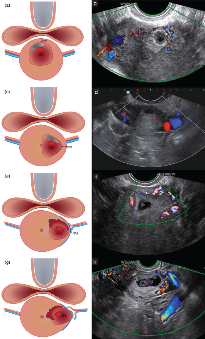
Figure 7.

Schematic (a,c,e,g) and ultrasound (b,d,f,h) images showing assessment of location of Cesarean scar pregnancy (CSP) in relation to the uterine arteries in the transverse plane. (a,b) Median location of CSP. (c,d) Eccentric location of CSP; the gestational sac (GS) is connected with the cervical canal and is within the outer cervical contour. (e,f) Lateral location of CSP; the GS protrudes towards the broad ligament within the virtual outer cervical contour and the residual myometrium is visible (CSP with largest part of GS embedded in the myometrium and not crossing the serosal line). (g,h) Lateral location of CSP; the GS is bulging beyond the outer cervical contour and residual myometrium is absent (CSP crossing the serosal line). RMT, residual myometrial thickness.
