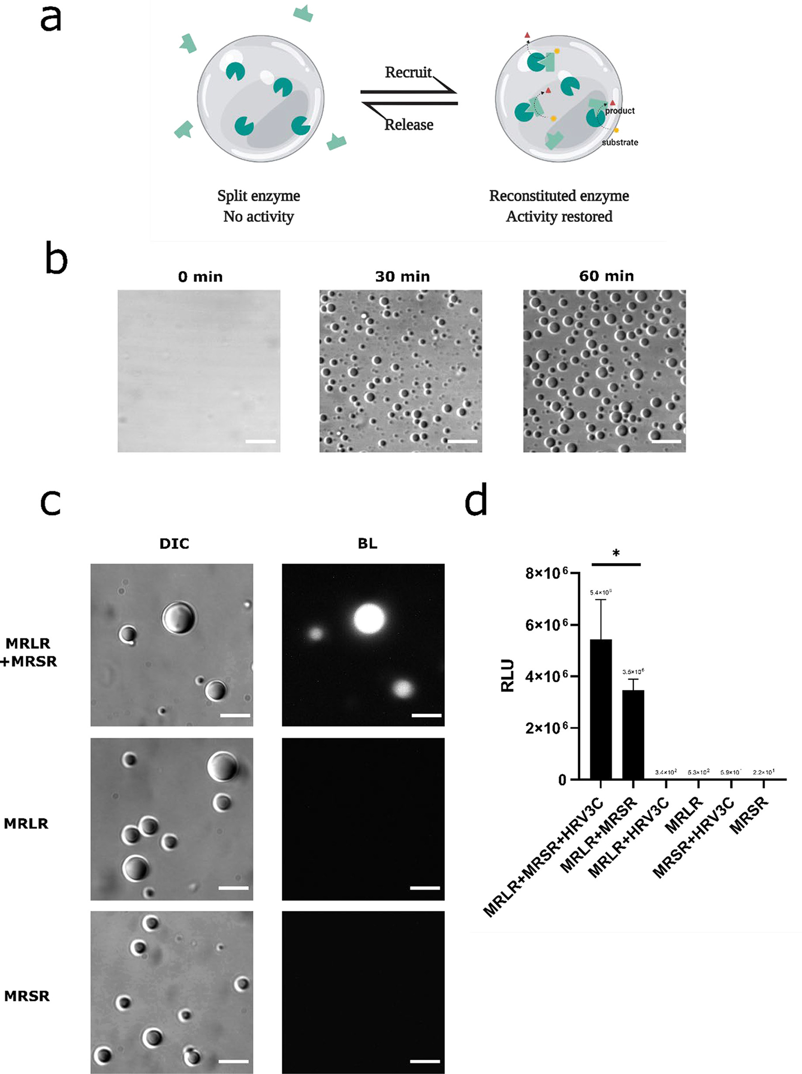
Figure 6.

Incorporation of fragmented Nanoluc enzyme into the RGG protein coacervates. (a) design schematic: Formation of RGG protein coacervates promotes reconstitution of the split enzyme and turns on biochemical reaction inside the coacervates. (b) RGG-LgBit-RGG and RGG-SmBit-RGG form coacervates. HRV3C protease (1μM final concentration) was added into a mixture of 3 μM MBP-x-RGG-LgBit-RGG and 3 μM MBP-x-RGG-SmBit-RGG (‘x’ denotes for HRV3C cleavage site) to cleave off the MBP domain and promote phase separation. Images of droplets were taken at different time points after protease addition. Scale bars: 10 μm. (c) Bioluminescence images showing reconstitution of LgBit and SmBit inside RGG protein coacervates. Top row: DIC and Bioluminescence images of protein droplets taken 1hr after adding HRV3C protease (1 μM final concentration) into a solution containing 3 μM MBP-x-RGG-LgBiT-RGG (abbreviated as ‘MRLR’) with 3 μM MBP-x-RGG-SmBiT-RGG (abbreviated as ‘MRSR’, ‘x’ denotes for HRV3C cleavage site), confirming reconstitution of NanoLuc activity from LgBit and SmBit inside the RGG-based coacervates. Middle row: DIC and Bioluminescence images of protein droplets taken 1 hr after adding HRV3C protease (1 μM final concentration) into a solution containing 6 μM MBP-x-RGG-LgBiT-RGG, indicating LgBit alone does not generate a light signal inside RGG-based droplets. Bottom row: DIC and Bioluminescence images of protein droplets taken 1hr after adding HRV3C protease (1 μM final concentration) into a solution containing 6 μM MBP-x-RGG-SmBiT-RGG, indicating SmBit alone does not generate a light signal inside RGG-based droplets. Scale bars: 10 μm. Substrate: 10 μl. Exposure: 5 s. (d) Light intensity readings from a plate reader for samples containing single or both components of NanoBit. From left to right: MBP-RGG-LgBit-RGG (abbreviated as ‘MRLR’) (3 μM) + MBP-RGG-SmBit-RGG (abbreviated as ‘MRSR’) (3 μM) + HRV3C (2 μM); MBP-RGG-LgBit-RGG (3 μM) + MBP-RGG-SmBit-RGG (3 μM); MBP-RGG-LgBit-RGG (6 μM) + HRV3C (2 μM); MBP-RGG-LgBit-RGG (6 μM); MBP-RGG-SmBit-RGG (6 μM) + HRV3C (2 μM); MBP-RGG-SmBit-RGG (6 μM). 5 μL of 200x diluted Nano-Glo assay substrate was injected into each well. Data presented as mean ± SD. *, p < 0.05; 2-tailed t-test.
