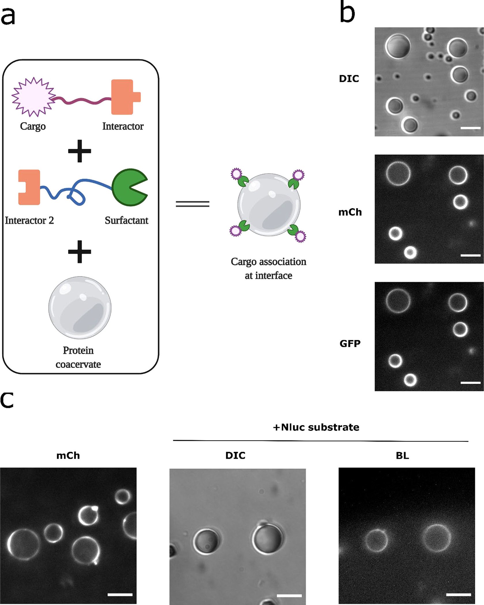
Figure 7.

Recruitment of NanoLuc enzyme at coacervate-water interface. (a) Schematic of design strategy: linking the cargo via interactor modules onto a surfactant molecule that localizes at the coacervate-water interface. (b) Recruitment of a sample cargo protein. SZ1-GST-mCherry-RGG and GFP-SZ2 were mixed at a ratio of 2:1 and incubated for 1 hr to optimize binding, then 2 μl of the mixture was titrated into 50 ul of protein solution containing 6 μM RGG-RGG, 150 mM NaCl. Final concentrations for SZ1-GST-mCherry-RGG, GFP-SZ2, and RGG-RGG were about 200 nM, 100 nM and 6 μM, respectively. From top to bottom: DIC image (exposure: 100 ms) on the droplets, mCherry channel (exposure: 100 ms) showing surfactant localization at the droplet interface, GFP channel (exposure: 1000 ms) showing localization of the GFP cargo at the coacervate interface. Scale bars: 10 μm. (c) Recruitment of NanoLuc enzyme at the coacervate interface. SZ1-GST-mCherry-RGG and NanoLuc-SZ2 were mixed and incubated, then titrated into protein solution containing 6 μM RGG-RGG, 150 mM NaCl. Final concentrations for SZ1-GST-mCherry-RGG, NanoLuc-SZ2, and RGG-RGG were about 800 nM, 400 nM and 6 μM, respectively. Left: mCherry image showing surfactant localization at the droplet interface, imaged using a spinning disk confocal to minimize out-of-focus light. Middle to right: DIC and Bioluminescence images showing NanoLuc recruitment at the coacervate-water interface, after addition of 10 μL Nano-Glo substrate. Exposure for bioluminescence imaging: 10 s. Scale bars: 10 μm.
