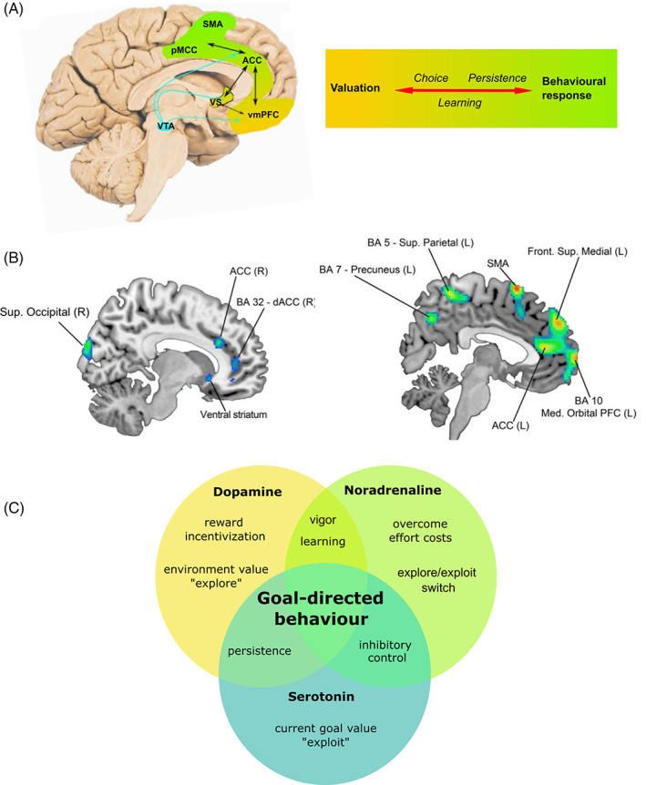
FIG 2.

Neurobiology of goal‐directed behavior. (A) Brain regions forming a network that underlies all phases of goal‐directed behavior. Color shading from gold to green represents transition from valuation neural regions and cognitive processes to motor regions and processes, leading to action. (B) Structural (L) and metabolic (R) correlates of apathy in HD. Key frontostriatal regions subserving goal‐directed behavior are implicated, notably the medial prefrontal cortex, anterior cingulate cortex, and ventral striatum. (C) Crucial neuromodulators implicated in goal‐directed behavior. Although simplex sigillum veri (simplicity is a sign of truth), this simplistic Venn diagram does not capture the complexities of goal‐directed behavior neuromodulation. Here we identify the predominant neuromodulatory system driving each cognitive process based on current animal models and human studies of goal‐directed behavior, bearing in mind their multiplex interdependence. Identification of the specific cognitive processes that are disrupted in apathy and/or impulsivity is key to developing therapeutic interventions. ACC, anterior cingulate cortex; BA, Brodmann area; dACC, dorsal anterior cingulate cortex; L, left; PFC, prefrontal cortex; pMCC, posterior midcingulate cortex; R, right; SMA, supplementary motor area; vmPFC, ventromedial prefrontal cortex; VTA, ventral tegmental area; VS, ventral striatum. (A) Adapted with permission from Le Heron et al. 49 (B) Reprinted with permission from Martínez‐Horta et al. 50 [Color figure can be viewed at wileyonlinelibrary.com]
