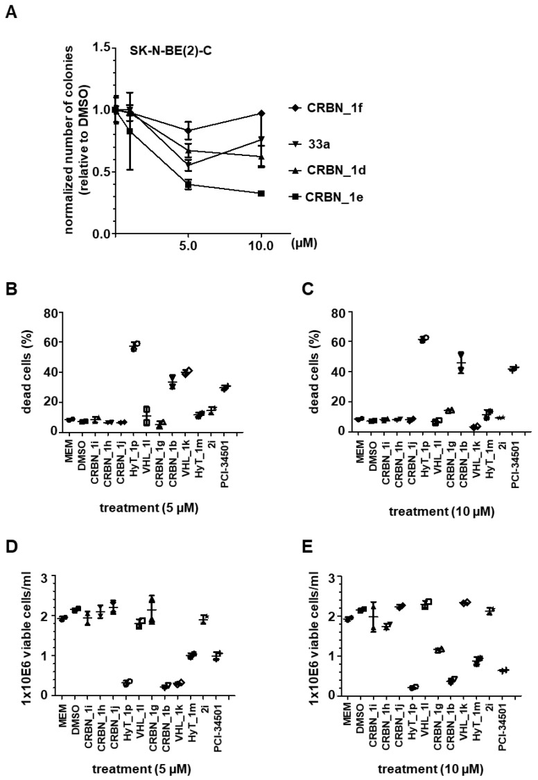
Figure 5.
(A) Neuroblastoma SK-N-BE(2)-C cells; Colony Assay, 10 days (compound treatment within first 96 h). Stained with crystal violet and quantified with ImageJ. (B,C) Trypan Blue assay for detection of dead cells. Neuroblastoma IMR-32 cells were either treated with 5 µM (B) or 10 µM (C). (D,E) Cell proliferation assessed via counting of viable cells. IMR-32 cells were either treated with 5 µM (D) or 10 µM (E) PROTACs. HDAC8i PCI-34501 served as a positive control, untreated (MEM) and solvent (DMSO) treated cells served as negative controls.

