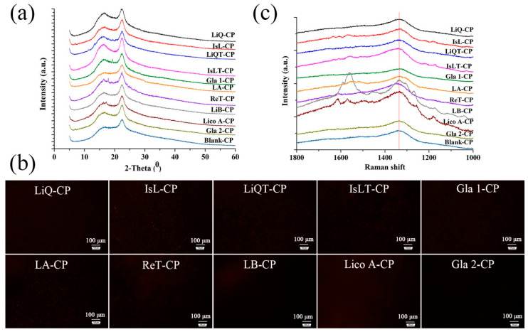
Figure 1.
Characterization of LFs-CP hydrogel. (a) X-ray powder diffractograms of ten LFs-CP hydrogels; (b) PLM images of LFs-CP films (The black represented that the drug was molecularly dispersed in the hydrogel films while red represented the crystals. Bar = 100 μm); (c) Raman spectra of different hydrogels.

