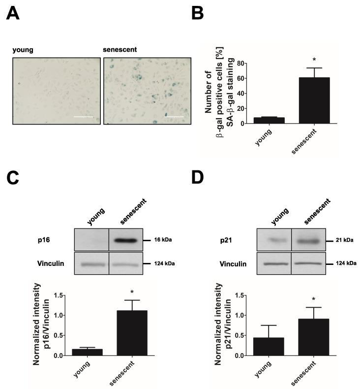
Figure 1.
Replicative senescence in endothelial cells. HUVEC of passage 1 (young) and passage 20–25 (senescent) were subjected to analysis of senescence markers. (A) Senescence-associated-β-galactosidase (SA-β-gal) staining. Representative picture out of n = 3, scale bar = 200 µm. (B) Quantification of SA-β-gal-positive cells (n = 3, * p < 0.05, unpaired Student’s t-test with Welch’s correction). (C,D) Representative western blots of senescence markers p16 and p21 (upper panels) and densitometric quantification of p16 and p21 after normalization to vinculin (lower panels, n = 5, * p < 0.05, for p16: unpaired Student’s t-test with Welch’s correction; for p21: unpaired Student’s t-test). All data represent mean + SD.

