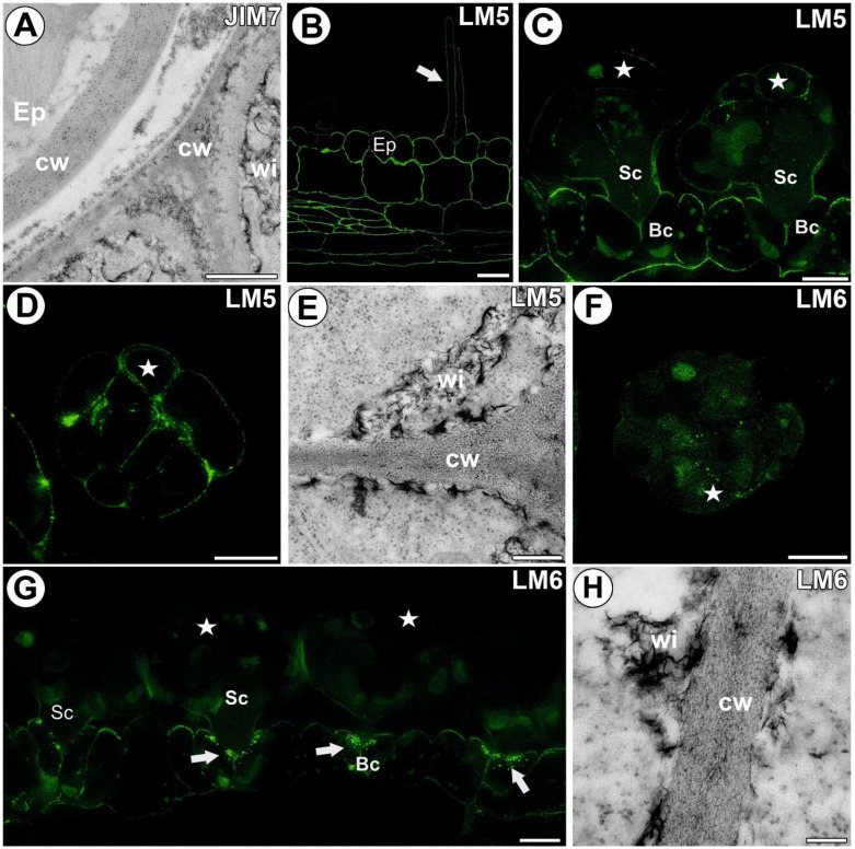
Figure 4.
Homogalacturonans detected in the A. vesiculosa trap. (A) Immunogold labeling of the cell wall with JIM19 in the ordinary epidermal (Ep) and glandular cells; wall ingrowths (wi), cell wall (cw); bar 900 nm. (B) HG (labeled with LM5) detected in the trap wall; epidermal cell (Ep), sensory trichome (white arrow), bar 10 µm. (C) HG (labeled with LM5) detected in the glands; secretory cell (star), stalk cell (Sc), basal cell (Bc), bar 10 µm. (D) HG (labeled with LM5) detected in the gland, paradermal section; secretory cell (star), bar 10 µm. (E) Immunogold labeling of the wall ingrowths with LM5 in the glandular cells; wall ingrowths (wi), cell wall (cw); bar 300 nm. (F) HG (labeled with LM6) detected in the gland, transverse section; secretory cell (star), bar 10 µm. (G) HG (labeled with LM6) detected in the glands and trap wall; secretory cell (star), stalk cell (Sc), basal cell (Bc), wall labyrinth in the basal cell (white arrow), bar 10 µm. (H) Immunogold labeling of the wall ingrowths with LM6 in the glandular cells; wall ingrowths (wi), cell wall (cw); bar 100 nm.

