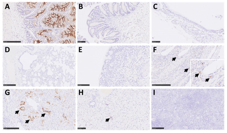
Figure 7.
Representative images of IHC for ACE2 receptor in marmoset tissues. Positive staining was observed in the small intestine (Bar = 250 µm) (A), mostly in the apical border of absorptive epithelial cells and at a low level in some epithelial cells of glands. Minimal staining in epithelial cells from the large intestine (Bar = 100 µm) (B). The positive staining in the trachea (Bar = 100 µm) (C), lung (Bar = 250 µm) (D), and respiratory nasal mucosa (Bar = 100 µm) (E) was almost non-existent. However, moderate positive staining was observed in glandular epithelium within the olfactory mucosa area of the nasal cavity (arrows and insert; Bar = 250 µm) (F). To the contrary, there was no staining in the epithelium lining or nerve terminations. Intense staining was observed in the kidney in the apical border of kidney tubular epithelial cells (arrows; Bar = 250 µm) (G) and moderate reaction in immune cells within the liver (Bar = 100 µm; arrows, normally extramedullary haematopoiesis) (H). No reaction was observed in the spleen (Bar = 250 µm) (I).

