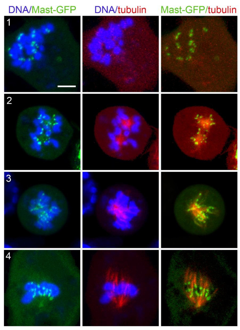
Figure 10.
Mast-GFP localization during kinetochore-driven microtubule regrowth (KDMTR) after colcemid-induced MT depolymerization. Live S2 cells expressing Mast-GFP (green) and Cherry-tubulin (red) were stained with the vital DNA stain Hoechst 33342 (blue). Mast-GFP is associated with discrete sites on the chromosomes (probably corresponding to the kinetochores) after 3 h colcemid treatment, before washout of the drug (panel (1)). During KDMTR, Mast-GFP is still accumulated on the kinetochores, which surround tubulin clusters/asters (likely mini spindle poles) that exhibit weak GFP fluorescence (panel (2)). Mast-GFP remains concentrated on kinetochores during further KDMTR stages (panels (3,4)). Scale bar, 5 µm.

