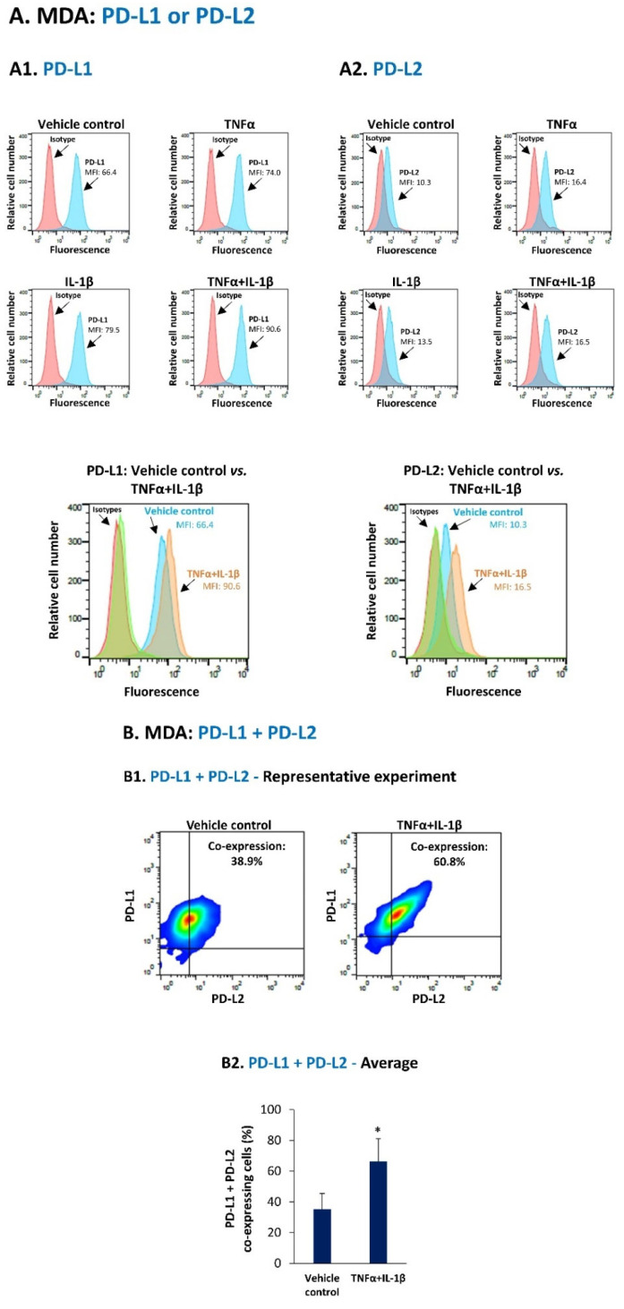
Figure 1.
Joint stimulation by TNFα + IL-1β promotes the proportion of PD-L1 + PD-L2 co-expressing MDA-MB-231 cells. (A) MDA-MB-231 cells (MDA) were stimulated by TNFα and/or IL-β (TNFα: 50 ng/mL; IL-1β: 500 pg/mL) for 24 h. Control cells were treated by the vehicle of the cytokines. Cytokine concentrations were selected as described in Section 2. Cell surface expression of PD-L1 (A1) and PD-L2 (A2) was determined by flow cytometry; MFI, mean fluorescence intensity. Isotype/s, Non-relevant isotype-matched antibodies, used as control/s. A representative experiment of n = 3 is presented. (B) MDA cells were stimulated by TNFα + IL-1β or vehicle, as in Part A. The proportion of cells co-expressing PD-L1 + PD-L2 was determined by flow cytometry. Axes determining PD-L1 + PD-L2 positive cells were set based on isotype staining; The percentages of PD-L1 + PD-L2 co-expressing cells were calculated by subtracting the percentages of PD-L1 + PD-L2 co-expressing cells in isotype-labeled cells from the percentages of PD-L1 + PD-L2 co-expressing cells, following staining by specific antibodies to PD-L1 and PD-L2. (B1) A representative experiment of n = 3 is presented. (B2) Average ± SD of n = 3 experiments is presented. * p < 0.05. Statistical analyses were performed as described in Section 2.

