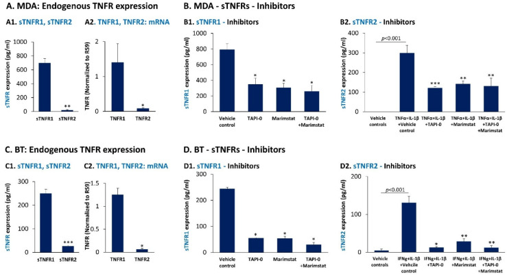
Figure 6.
MDA-MB-231 and BT-549 cells express higher levels of cell-derived sTNFR1 than sTNFR2, and both soluble receptors are shed in an ADAM17-dependent process. (A,C) The constitutive cell-derived expression levels of sTNFR1 and sTNFR2 were determined by ELISA in CM of MDA-MB-231 cells (MDA) (A1) and BT-549 cells (BT) (C1), after 48 h of cell growth. In parallel, mRNA levels of TNFR1 and TNFR2 were determined by qPCR in MDA cells (A2) and BT cells (C2), after 5 h of cell growth. In each panel, a representative experiment of n = 3 is presented. *** p < 0.001, ** p < 0.01, * p < 0.05. (B,D) Cell-derived levels of sTNFR1 (B1,D1) and sTNFR2 (B2,D2) were determined in a 48 h CM of MDA cells (B1,B2) and in BT cells (D1,D2) following treatment with marimastat (3.3 µg/mL), TAPI-0 (5 µg/mL) and both inhibitors together (same concentrations), as indicated. Inhibitor concentrations were selected as described in Section 2 and they did not affect tumor cell growth. To enable determination of the inhibitors on sTNFR2 levels, the experiments in Parts B2 and D2 were performed in the presence of cytokine stimulation: TNFα + IL-1β for MDA cells and IFNγ + IL-1β for BT cells (cytokine concentrations were as in Figure 1 and Figure 2, respectively). Control cells were treated by the vehicle of the inhibitors and/or of the cytokines. In each panel, a representative experiment of n = 3 is presented. *** p < 0.001, ** p < 0.01, * p < 0.05. Statistical analyses were performed as described in Section 2.

