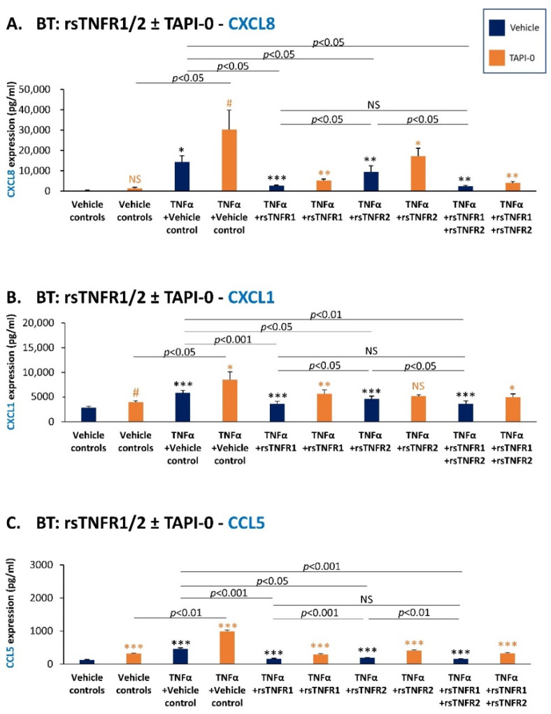
Figure 10.
Recombinant and cell-derived sTNFR1 and sTNFR2 have a protective effect against TNFα-induced expression of pro-metastatic chemokines in BT-549 cells. BT-549 cells (BT) were stimulated by TNFα (0.25–0.5 ng/mL) that was pre-incubated with rsTNFR1 (150 ng/mL), rsTNFR2 (500 ng/mL), rsTNFR1 + rsTNFR2 (concentrations as before) or their vehicle. When indicated, the cells were cultured prior to TNFα stimulation with TAPI-0 (5 µg/mL) or its vehicle for 3 h, as well as during cytokine stimulation (TAPI-0 did not affect tumor cell growth). The concentrations of rsTNFR1 and rsTNFR2 were selected as described in Section 2. CM were collected following 24 h stimulation, and CXCL8 (A), CXCL1 (B) and CCL5 (C) levels were determined by ELISA. A representative experiment of n = 3 is presented. *** p < 0.001, ** p < 0.01, * p < 0.05, # p < 0.1. NS, not significant. Black asterisks denote the differences in chemokine levels between TNFα-stimulated cells and vehicle-treated cells. Orange asterisks denote the differences in chemokine levels between TAPI-0-treated cells and cells treated by its vehicle. Statistical analyses were performed as described in Section 2.

