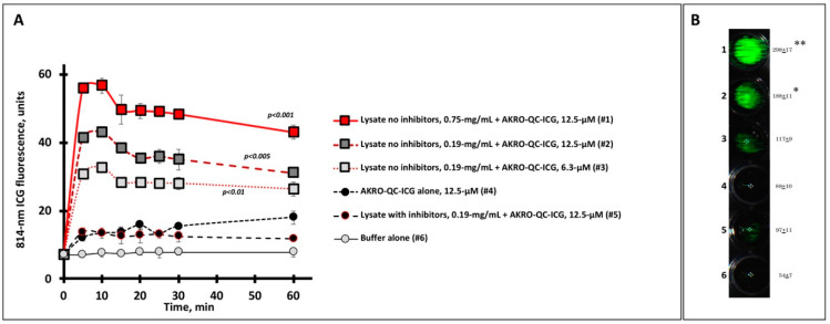
Figure 2.
Human BCa cell lysate initiates specific fluorescence imaging of AKRO-QC-ICG probe in vitro. (A)—Time course of ICG fluorescence imaging of cancer-cell-associated cysteine cathepsins (lysate) mixed with substrate (AKRO-QC-ICG). Tecan fluorescent scanner was used to assess ICG fluorescence in 4 wells per sample for different mixes, probe alone, or buffer alone (100 μL per well, 96-well black flat bottom plate) in three experiments. Irreversible cysteine protease inhibitor K777 was used as a control for specificity of fluorescence. Vertical bars—SD. (B)—Representative sample of ICG FL in AKRO-QC-ICG/MDA-MB-468 cancer cell lysate mixes after 10 min incubation. Data presented as mean fluorescence ± SD in 3 wells per sample. Number of wells corresponds to the number of samples tested in (A). Quantification of well signal in arbitrary units after 10 min of incubation is presented to the right of each well. Imaging of lysate spots was performed using the Curadel Lab-Flare RP1 with 800 nm filter set and Curadel Resvet Imaging software. Notes: (1) the levels of fluorescence of all lysates without inhibitor were significantly higher as compared to controls (wells #4–#6) in (A); *—p < 0.05, **—p < 0.01 compared to controls (wells #4–#6) in (B) by Student’s t-test in three independent experiments both for (A) and (B)).

