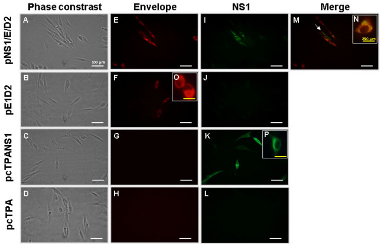
Figure 2.
In vitro expression of the E and NS1 proteins in cells transfected with the different DNA vaccines. BHK-21 cells were transiently transfected with pNS1/E/D2, pE1D2, pcTPANS1, or the negative control pcTPA. The E protein was detected with the mouse 3H5 monoclonal antibody (against domain III) and goat anti-mouse IgG Alexa Fluor 546 antibody (red fluorescence). The NS1 protein was detected with rabbit anti-NS1 polyclonal serum, followed by incubation with goat anti-rabbit IgG Alexa Fluor 488 antibody (green fluorescence). Phase contrast (A–D). Cells were incubated with anti-EDIII of the E protein (E–H,O) and anti-NS1 (I–L,P) antibodies. Images merge (M,N). Cells were analyzed by fluorescence microscopy at 40× (A–M) or 100× (N–P) magnifications. The white arrow indicates the pNS1/E/D2-transfected cell expressing only the NS1 protein. Scale white bar, 100 μm; scale yellow bar, 250 μm.

