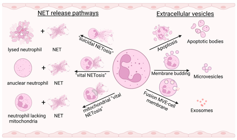
Figure 1.
Mechanisms of NET formation (left) and EV classes released by neutrophils (right). In “suicidal NETosis”, chromatin is released from the lysing neutrophil after chromatin de-condensation and subsequent rupture of the nuclear and cell membrane. “Vital NETosis” is characterized by the fusion of vesicles containing nuclear DNA with the plasma membrane, finally resulting in an anuclear cytoplast. In a second form of “vital NETosis”, neutrophils release mitochondrial DNA by an unknown NOX-dependent mechanism. Extracellular vesicles comprise a group of heterogeneous membranous vesicles of varying size and morphology. Apoptotic bodies represent subcellular fragments after the disassembly of a dying cell. The smaller microvesicles bud from the cell membrane and contain cytoplasmic material. The smallest EVs are referred to as exosomes and are released from the lumen of multivesicular endosomes (MVE), fusing with the cell membrane. Created with BioRender.com.

