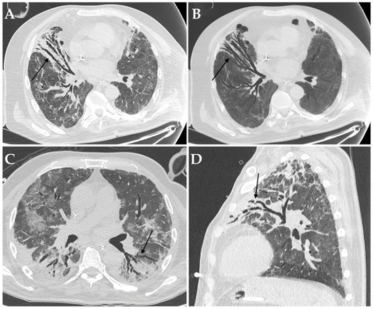
Figure 4.
Axial HRCT images of a patient with COVID-19 ARDS admitted to ICU with bronchiectasis in the middle lobe (black arrow in (A)), being more evident at Minimum Intensity Projection (MIP) reconstruction (black arrow in (B)); Aspergillus niger was later detected in the bronchoalveolar lavage of the patient. Axial HRCT image of a different patient with COVID-19 ARDS with superinfection by Klebsiella pneumoniae and bronchiectasis in the dorsal segment of the left upper lobe (black arrow in (C)). Sagittal HRCT image of a different patient with COVID-19 ARDS showing bronchiectasis in the left upper lobe (black arrow in (D)) and presenting with elevated serum-galactomannan levels compatible with Aspergillus spp. superinfection.

